The secret social network hiding inside your fascia
Why Carla Stecco and colleagues' new discovery is so exciting for movement teachers and manual therapists
For years, we've been looking at fascia rather like the physical infrastructure of a city.
We've studied its collagen fibres, elastic networks and the remarkable gel-like substance, hyaluronan, that helps create its hydrated extracellular environment.
We've discovered cells that manufacture and remodel this environment. We've learned that fascia contains nerves, blood vessels, lymphatics and immune cells.
But now, researchers have identified something crucial about the coordination of all this... stuff.
A network of cells that may help connect some of these systems together.
They're called telocytes.
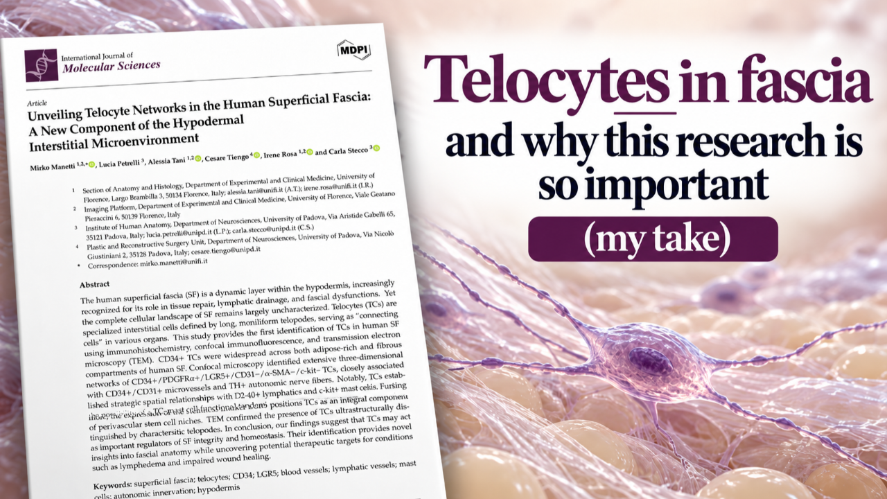
And a new paper by Mirko Manetti, Carla Stecco and colleagues, published on 20 October 2026, has mapped these extraordinary cells throughout the human superficial fascia for the first time using a combination of microscopy techniques.
Meet the telocytes: the neighbourhood connectors
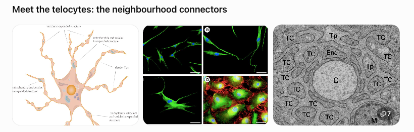
Imagine tiny neighbourhood coordinators with extraordinarily long arms.
Telocytes have relatively small cell bodies, but they extend delicate, thread-like projections called telopodes through the surrounding connective tissue.
These projections are so fine that specialized microscopy is needed to appreciate their architecture.
And rather than sitting in isolation, the cells form elaborate three-dimensional networks.
Think of a neighbourhood WhatsApp group — except instead of people sending messages between houses, we're looking at cells potentially exchanging information within a living tissue.
The exciting part isn't simply that these cells exist. Telocytes have already been identified in other organs.
It's where the researchers found them.
Throughout the superficial fascia, these cells were positioned close to:
-
Blood vessels: the supply routes bringing oxygen and nutrients.
-
Lymphatic vessels: the drainage routes involved in clearing excess interstitial fluid.
-
Mast cells: immune cells that can release powerful chemical messengers.
-
Sympathetic nerve fibres: part of the autonomic nervous system, involved in regulating vascular responses.
-
Potential regenerative niches: environments containing cells involved in tissue maintenance and repair.
This arrangement raises a fascinating possibility: telocytes might help coordinate the activities of several systems we have traditionally studied separately.
Why this matters when you move or touch someone
Imagine a sponge woven with elastic threads.
The sponge isn't simply wet or dry. Its behaviour depends on the arrangement of its fibres, the composition of its gel, the movement of fluid and the activity of the cells living within it.
Now imagine that some of those cells can detect changes in their surroundings and potentially communicate with neighbouring cells.
Suddenly, the sponge isn't such a passive material anymore.
This is the exciting direction in which fascial science is moving.
When you stretch an arm overhead, walk, squat or receive manual therapy, you're applying mechanical forces to living tissues containing all these interacting systems.
The question becomes more interesting than simply:
How much did the fascia stretch?
We can also ask:
How might the cells within this tissue sense, communicate and adapt to the changing mechanical environment?
The new paper doesn't demonstrate that telocytes respond to a yoga pose, a massage technique or a particular exercise. But it identifies an anatomical network that makes those questions worth investigating.
Here's where the story gets even more exciting: the gel
This is the connection I find especially intriguing.
We've already been exploring the role of hyaluronan (HA) — the water-associated molecule that contributes to the physical environment surrounding fascial cells.
Think of collagen as part of the woven architecture of our city, and hyaluronan as a major ingredient of the hydrated environment filling its spaces.
HA isn't simply a lubricant. Its molecular size, concentration, organization and interactions with receptors such as CD44 and RHAMM can influence cellular behaviour.
My work with collaborators explores how calcium signalling and HA metabolism participate in feedback between cells and their extracellular environment.
Now Stecco and colleagues have revealed an extensive network of telocytes occupying this same interstitial territory.
Could these cells help sense changes in the hydrated matrix? Could they communicate with immune cells when the local environment changes? Could they contribute to the coordination of tissue repair?
These are exciting questions because they bring together three important elements of fascial biology:
- The matrix — collagen, elastin, hyaluronan and water create the physical environment.
- The cellular network — telocytes may help organize local interactions and communication.
- The response — tissue maintenance, inflammation, fluid regulation and repair may emerge from interactions between these systems.
But there's an important distinction: this new study did not measure HA, CD44, RHAMM or calcium signalling in the identified telocytes. The proposed molecular connection remains a hypothesis, not a demonstrated mechanism.
Rich clue: the lymphatic connection
Here's where our city analogy becomes particularly interesting.
Imagine a neighbourhood experiencing a sudden flood.
It needs some way of detecting the change in fluid pressure and coordinating a response from its drainage system.
The authors discuss recent research in mouse joints suggesting that telocytes can participate in a remarkable sequence:
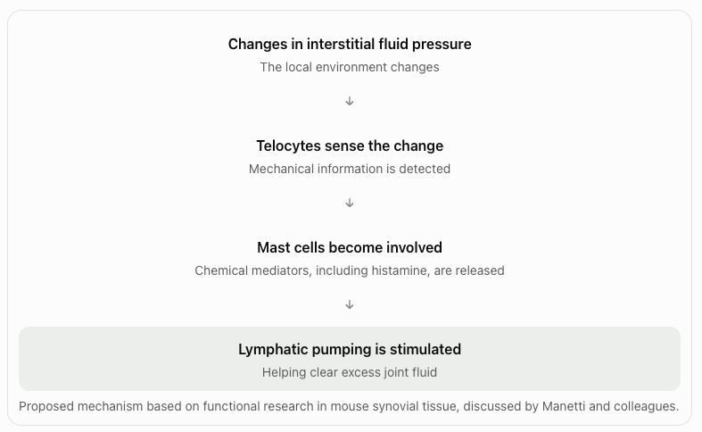
And now, in human superficial fascia, the researchers have found telocytes sitting extraordinarily close to both lymphatic vessels and mast cells.
It's rather like discovering that the neighbourhood coordinators are stationed right beside the drainage infrastructure and its chemical-response teams.
We don't yet know whether they perform the same function in human superficial fascia. But we now have an anatomical reason to investigate it.
What does this change for movement teachers and manual therapists?
For me, it reinforces an important shift in how we understand living tissue.
We cannot reduce fascia to something that needs to be pulled longer, broken apart or mechanically released.
It is an active, cellular, hydrated environment, continually maintaining itself and responding to changing conditions.
This paper gives us another piece of that picture.
It encourages us to think about movement and touch not simply as forces acting upon material, but as mechanical inputs experienced by living cells embedded within a responsive matrix.
That doesn't mean we've proven that a particular technique activates telocytes or improves lymphatic drainage. We haven't.
The study examined abdominal superficial fascia from just three donors, and the authors themselves call for functional research, including experiments investigating mechanical loading, physical therapy and manual interventions.
But it offers a compelling new direction for understanding how tissues might coordinate their responses to movement, inflammation, fluid changes and injury.
And that's why I find this discovery so exciting.
We're beginning to move beyond anatomy as a map of structures, towards anatomy as an ecology of relationships.
The fibres - The gel - The cells all matter.
And perhaps, increasingly, what matters most is the conversation happening between them.
That's a much more interesting way to think about fascia — and an exciting scientific foundation for continuing to explore Glide Before Gains.
Research discussed: Manetti, M., Petrelli, L., Tani, A., Tiengo, C., Rosa, I., & Stecco, C. (2026). Unveiling Telocyte Networks in the Human Superficial Fascia: A New Component of the Hypodermal Interstitial Microenvironment. International Journal of Molecular Sciences, 27(20), 9331. Read the open-access paper .

YOUR FREE GLIDE GUIDE
Stiffer, puffier or simply less at ease in your body lately? Discover how oestrogen, progesterone, relaxin and cortisol shape the way your fascia feels, and how to move with your hormones, not against them.