What do Egyptian mummies have to do with women’s fascia, collagen and menopause? They offer a striking way to think about collagen’s durability — and how different living tissue is from tissue preserved after death.
This blog was inspired by David's comment on my Facebook page where I posted my blog on whether it's worth retiring the term "fascia".

So how did the Ancient Egyptians encounter fascia, and what is known here? The bridge is collagen: the structural protein that forms much of connective tissue, from skin and bone to the sheaths around muscles.
It is a fair question.
At first glance, dragging four-thousand-year-old linen-wrapped bodies into a contemporary nomenclature debate feels a bit like bringing a sarcophagus to a biomechanics symposium.
But the short, unvarnished answer to David’s question is simple and it's all Hyp.
No, not hype in the buzzword sense—though heaven knows the fascia community has seen plenty of that.
We're talking Hyp: hydroxyproline, the non-proteinogenic amino acid workhorse that stabilizes the collagen triple helix, defies enzymatic degradation, and allows soft tissues to survive centuries of desiccation, resins, and shifting scientific paradigms.
When we debate whether to keep, split, or retire the word “fascia,” we are ultimately wrestling with how to describe the body’s continuous, collagenous matrix.
And if you want to understand what makes that matrix chemically unique, physically durable, and anatomically stubborn, you have to look at what happens when human tissue is pushed through the ultimate preservation test.
One of collagen’s molecular stabilisers is hydroxyproline, or Hyp. Its chemistry helps collagen chains hold their triple-helix shape. Collagen-rich tissues can leave a durable trace — even after mummification has radically changed their water content and form.
Estrogen does not directly make hydroxyproline by switching on the prolyl-hydroxylase reaction. Its connection is broader: research suggests estrogen can support maintenance of collagen-rich tissues and limit some pathways of matrix breakdown.
From proline to a resilient collagen helix
Hydroxyproline is made when enzymes modify selected proline residues during collagen production; it is not simply inserted intact from food into newly built collagen.
within a collagen chain → Prolyl hydroxylase
O2 · iron · ascorbate → Hydroxyproline
triple-helix stability
1. 2500 BC: The Ultimate Practical Soft-Tissue Workshop
Let’s be completely clear about one thing: ancient Egyptian embalmers were not reading modern anatomical terminology or philosophizing about tensegrity.
They did not have the formal theoretical taxonomy that later developed in classical Greece or the Alexandrian medical school of the 4th century BC (Toprak et al., 2025).
What they did have was centuries of sustained, tactile, hands-on encounters with human tissue compartments, organ membranes, and structural body walls (Toprak et al., 2025).
The roots of this practice stretch back far further than most realize.
Chemical analysis of funerary wrappings from Late Neolithic burials (c. 4500–3350 BC) at Mostagedda in Upper Egypt demonstrates that complex embalming recipes—blending plant oils, animal fats, conifer resins, aromatic extracts, and waxes with natural antibacterial properties—were already in use millennia before the pharaohs built the pyramids (Jones et al., 2014).
By the Old Kingdom (c. 2670–2500 BC), embalming matured into a sophisticated ritual technology featuring canopic jars dedicated to specific viscera and individual limb-wrapping techniques designed to preserve the body's anthropomorphic form (Jones et al., 2014; Öhrström et al., 2020; Zesch et al., 2024).
| Period | Date | Embalming practice | Reference |
|---|---|---|---|
| Late Neolithic | c. 4500–3350 BC | Linen wrappings impregnated with resin and antibacterial recipes | Jones et al., 2014 |
| Old Kingdom | c. 2500 BC | True mummification begins; canopic jars and compartmentalized evisceration | Öhrström et al., 2020; Zesch et al., 2024 |
| New Kingdom | c. 1300–1450 BC | Complex balms including beeswax, bitumen, Pinaceae and Pistacia resins | Huber et al., 2023 |
To execute evisceration, excerebration, and thorough desiccation with natron salts, embalmers had to systematically navigate skin layers, visceral investments, and fascial envelopes (Öhrström et al., 2020; Huber et al., 2023).
Although classical historians such as Herodotus and Diodorus Siculus documented these rituals centuries later, archaeological biochemistry confirms that the finest soft-tissue preservation was achieved long before Herodotus ever set foot in Egypt (Buckley & Evershed, 2001).
2. What Stays, What Shrinks, and What Falls Apart
If you want to understand how connective tissues behave under extreme physiological stress, experimental mummification provides a fascinating window.
When modern researchers replicate Egyptian mummification protocols using human and porcine models, the natron salt bath systematically pulls out water, halting enzymatic putrefaction and leaving behind remarkably intact skin and skeletal muscle (Papageorgopoulou et al., 2015).
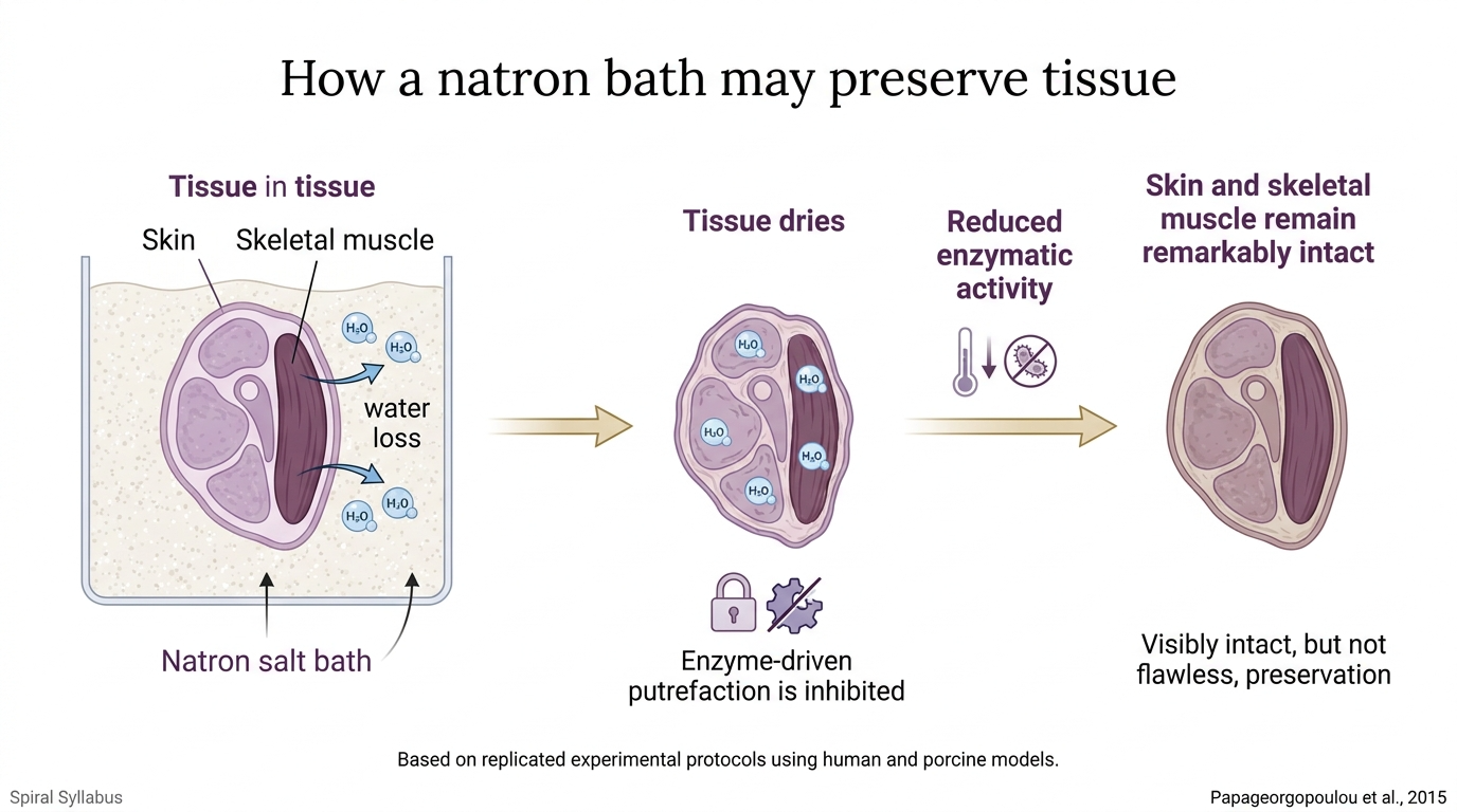
A 13-year experimental follow-up on porcine tissue confirmed that heart, muscle, and broad soft-tissue architectures can maintain their structural identity over extraordinary timeframes (Nerlich et al., 2025).
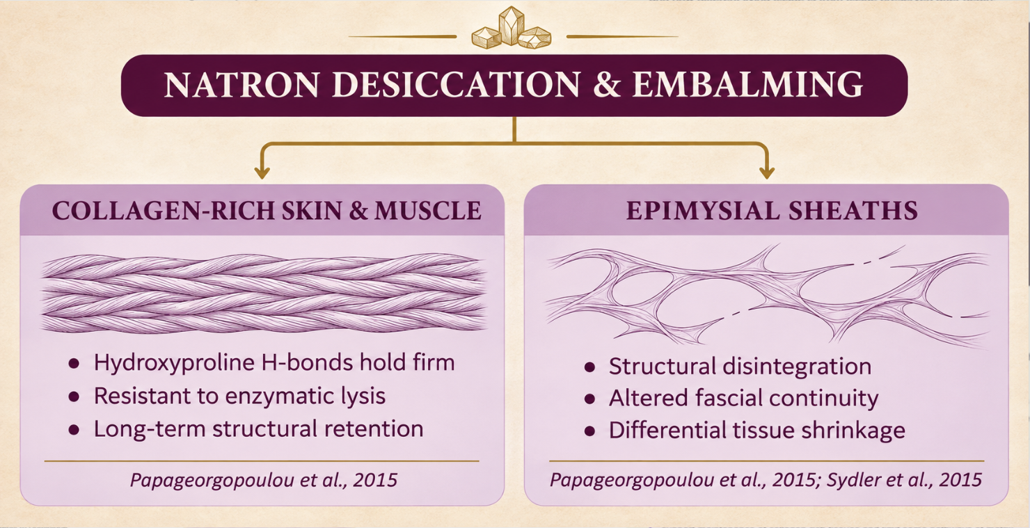
However, mummification is not a magic freeze-frame; it dramatically alters morphology:
-
Differential Shrinkage: Computed tomography (CT) comparisons across mummy types show that Egyptian mummies undergo marked volume loss compared to ice mummies, with distal extremities shrinking most aggressively due to high surface-area-to-volume ratios (Sydler et al., 2015). Interestingly, this shrinkage depends on tissue composition and preparation rather than how many millennia the mummy spent in a tomb (Sydler et al., 2015).
-
Fascial Disintegration: Under the microscope, the epimysium—the dense fascial layer encasing skeletal muscle bundles—undergoes significant structural disruption and breakdown during the mummification process, even while the primary muscle fibers and dermal layers survive (Papageorgopoulou et al., 2015).
Modern non-invasive analytics continuously expand our window into these altered tissues. High-resolution CT scans of individual mummies can trace residual soft-tissue planes, organ-removal trajectories, and ritual interventions (Goodman et al., 2026).
Meanwhile, electron microscopy of ancient dermis reveals surviving stratum corneum, keratinocytes, elastic fibers, and dense beds of collagen fibrils (Lewin, 1967; Zaki, 2023). Advanced organic residue analyses from 18th-dynasty canopic jar balms show intricate recipes of beeswax, plant oils, bitumen, and aromatic tree resins engineered to protect these exact tissue envelopes (Huber et al., 2023).
3. The Molecular Anchor: It’s All About Hyp
Why does the structural framework of the body outlast internal organs and cellular cytoplasm by four thousand years? Because that is prettymuch the upshot here.
Proteomic analysis of 4,200-year-old muscle and skin biopsies from First Intermediate Period mummies identified 230 unique proteins, with collagen emerging as overwhelmingly dominant (Jones et al., 2016).
The secret to collagen’s legendary survival is its primary stabilizing constituent: hydroxyproline (Hyp) (Definitions, 2020; Hu et al., 2021; Rappu et al., 2019).
The Biochemistry of Hydroxylation
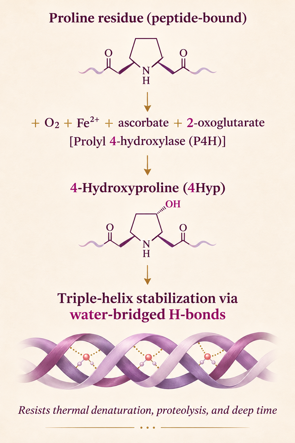
In vertebrate physiology, hydroxyproline is not incorporated directly from free dietary Hyp into newly forming polypeptide chains; rather, it is generated by co- and post-translational hydroxylation of peptide-bound proline residues (Kivirikko, 2024; Sadava & Chrispeels, 1971).
In simple, fun terms: your body does not just eat hydroxyproline and slot it intact into new collagen like a pre-assembled Lego brick.
Instead, think of collagen production like an artisan workshop weaving a heavy-duty biological rope.
First, standard proline molecules are loaded into the growing polypeptide chain. Then, specialized enzymes step in to give selected proline residues a chemical makeover—tacking on an oxygen and hydrogen group to turn them into hydroxyproline (Hyp).
That finished Hyp is the secret ingredient that lets the triple helix twist tightly and lock together into an ultra-stable, tear-resistant structure.
When biochemists describe this as “co- and post-translational,” they simply mean the makeover happens while the collagen chain is actively being built on the ribosome or immediately after it rolls off the line.
A side note on collagen supplements: This biochemical reality is precisely why we need to be critical (though not dismissive) about commercial collagen claims. Ingesting collagen hydrolysates does not act as an express delivery service sending intact dietary hydroxyproline straight into your tendon, skin, or fascia. Collagen supplements do supply amino acids and bioactive peptides that can signal cells and influence collagen turnover, but they are not plug-and-play tissue replacement parts. Real-world outcomes depend heavily on clinical evidence, therapeutic dose, product quality, and remembering that encouraging findings in animal models (like fish swim bladders!) do not automatically guarantee identical results in human connective tissues.
This crucial proline makeover is carried out by prolyl hydroxylases (P4H), which act as oxygen-dependent dioxygenases requiring four essential cofactors:
-
Molecular oxygen ($$\text{O}_2$$)
-
Ferrous iron ($$\text{Fe}^{2+}$$)
-
Ascorbate (Vitamin C)
-
2-oxoglutarate ($$\alpha$$-ketoglutarate)
The absolute requirement for oxygen makes this enzymatic system a direct biochemical oxygen sensor in animal biology (Journal of Biological Chemistry, 1963; Mócsai et al., 2021; Tiainen et al., 2005).
Structural Armor
Once formed, hydroxyproline confers extreme biomechanical stability:
-
Thermal and Conformational Resilience: Approximately half the proline residues in fibrillar collagen are converted to 4-hydroxyproline (4Hyp), which anchors the triple helix through water-bridged hydrogen bonding networks (Hudson et al., 2012). Molecular dynamics simulations confirm that inter-chain hydrogen-bond energy correlates directly with Hyp concentration; when Hyp content drops, thermal unfolding promptly begins (Xu et al., 2019).
-
Supramolecular Assembly: The rarer 3-hydroxyproline (3Hyp) participates in intermolecular cross-talk during fibril assembly; disrupting its specific enzyme complex causes severe recessive forms of osteogenesis imperfecta (Hudson et al., 2012).
-
Protease Resistance: The rigid pyrrolidine rings of proline and Hyp create sterically constrained peptide bonds that resist ordinary digestive and tissue proteases (Krane, 2008; Virgilio et al., 2024; Taga et al., 2022). Unmodified proline residues dictate matrix metalloproteinase (MMP) cleavage specificity, modulating turnover in living matrix (Krane, 2008).
Beyond Animals: A Universal Structural Motif
Nature likes this trick so much that hydroxyproline is by no means unique to animal connective tissues. Across the plant kingdom—from green algae and mosses to higher angiosperms—as well as in a range of fungi, Hyp residues form the backbone of hydroxyproline-rich glycoproteins (HRGPs), arabinogalactans, and extensins (Ashford & Neuberger, 1980; Johnson et al., 2017; Liang et al., 2020; Miller, 1971; Northcote, 1960; Rempfer et al., 2024).
In plants, Hyp is likewise formed by prolyl 4-hydroxylases utilizing ascorbate, $$\text{Fe}^{2+}$$, and $$\alpha$$-keto acids, and subsequent $$O$$-glycosylation provides cell-wall tensile strength, defense architecture, and developmental integrity (Kishor et al., 2015; Marcus et al., 1991; Petersen et al., 2021; Sadava & Chrispeels, 1971).
Furthermore, in plant cell walls, these Hyp-rich extracellular structures do not exist in a vacuum; they participate in dynamic signaling and remodeling networks that intersect with calcium-dependent regulation of cellular expansion, defense responses, and wall mechanics (Kishor et al., 2015; Marcus et al., 1991).
4. Supplements, Bioavailability, and the Broth Reality
In living systems, Hyp dynamics remain central to tissue remodeling, repair, and nutritional biochemistry:
-
Bioactive Signaling: When collagen hydrolysates are ingested, roughly 20–30% of total Hyp enters systemic circulation intact as small di- and tripeptides (such as Pro-Hyp, Hyp-Gly, and the exceptionally stable Gly-3Hyp-4Hyp) via intestinal peptide transporters (Taga et al., 2022; Virgilio et al., 2024). These circulating oligopeptides are not just passive nitrogen; they stimulate fibroblast chemotaxis, boost chondrocyte hyaluronic acid synthesis, and support osteoblast differentiation (Alves et al., 2023; Hsu et al., 2026; Lu et al., 2026; Salla et al., 2024; Zhu et al., 2025).
-
Dietary Precursors in Animal Models: In aquaculture and veterinary nutrition models, dietary Hyp supplementation directly upregulates collagen gene expression (col1a1, col1a2, p4h) and enhances structural matrix deposition (Cao et al., 2022; Guo et al., 2022, 2023; Li & Wu, 2017; Rong et al., 2019).
-
The Bone Broth Myth: While hydrolyzed collagen supplements deliver consistent, concentrated doses of Hyp peptides, ordinary bone broth is wildly variable. Standardized analyses reveal that commercial and homemade bone broths deliver substantially lower and unpredictable concentrations of hydroxyproline, proline, and glycine compared to measured supplement doses (Alcock et al., 2019).
Where estrogen enters the story
As estrogen changes through perimenopause and menopause, many women notice shifts in skin, joints and recovery. Older skin studies offer one window into the link between sex hormones and collagen.
A biopsy study found higher skin collagen and hydroxyproline among women treated with oestrogen-and-testosterone implants than in untreated, age-matched women (Brincat et al., 1983). Later work reported that some oestrogen regimens could prevent or restore aspects of postmenopausal dermal collagen loss (Brincat et al., 1987).
These small, older studies examined specific treatments. They do not establish that every modern HRT regimen has the same effect, that HRT is a treatment for fascia, or that a skin finding predicts how every connective tissue will respond. Menopausal Hormone Therapy (MHT) and Hormone Replacement Therapy (HRT) both use hormones (such as estrogen and progesterone) to manage hormonal changes, though medical organizations increasingly prefer the term MHT to describe menopause-specific treatments.
That is where the mummy analogy ends. A mummy can help us ask what physical structures persist; it cannot show how a living woman’s fascia feels, adapts or functions during menopause.
Living tissue is continually built, remodelled and repaired.
The useful question is not “Does estrogen make collagen?” It is “How might changing estrogen signalling influence collagen formation, retention and breakdown — and how does that differ by tissue and person?”
In the companion article, we follow that question through skin, bone and the cornea, separating what the studies measured from what we still cannot conclude about fascia.
References
Alcock, R. D., Shaw, G., & Burke, L. (2019). Bone broth unlikely to provide reliable concentrations of collagen precursors compared with supplemental sources of collagen used in collagen research. International Journal of Sport Nutrition and Exercise Metabolism, 29(3), 265–272. https://doi.org/10.1123/ijsnem.2018-0139
Alves, V., Gonçalves, S., Daguer, H., Micke, G., & Vitali, L. (2023). Development of a new method for the determination of 4-hydroxyproline as a measurement of collagen content in meat products and dietary supplements by cyclodextrin-assisted electrokinetic chromatography. Journal of Food Composition and Analysis, 124, 105431. https://doi.org/10.1016/j.jfca.2023.105431
Ashford, D., & Neuberger, A. (1980). 4-Hydroxy-L-proline in plant glycoproteins. Trends in Biochemical Sciences, 5(9), 245–248. https://doi.org/10.1016/s0968-0004(80)80813-9
Brincat, M., Moniz, C., Studd, J., Darby, A., Magos, A., & Cooper, D. (1983). Sex hormones and skin collagen content in postmenopausal women. British Medical Journal (Clinical Research ed.), 287, 1337–1338. https://doi.org/10.1136/bmj.287.6402.1337
Brincat, M., Versi, E., Moniz, C., Magos, A., De Trafford, J. C., & Studd, J. (1987). Skin collagen changes in postmenopausal women receiving different regimens of estrogen therapy. Obstetrics & Gynecology, 70, 123–127. https://doi.org/10.1016/0378-5122(88)90119-3
Buckley, S., & Evershed, R. (2001). Organic chemistry of embalming agents in Pharaonic and Graeco-Roman mummies. Nature, 413, 837–841. https://doi.org/10.1038/35101588
Cao, S., Xiao, Y., Huang, R., Zhao, D., Xu, W.-Q., Li, S., Tang, J.-Z., Qu, F.-F., Jin, J.-Y., Xie, S.-Q., & Liu, Z. (2022). Dietary supplementation with hydroxyproline enhances growth performance, collagen synthesis and muscle quality of Carassius auratus triploid. Frontiers in Physiology, 13, 913800. https://doi.org/10.3389/fphys.2022.913800
Cosman, F., Shen, V., Xie, F., Seibel, M., Ratcliffe, A., & Lindsay, R. (1993). Estrogen protection against bone resorbing effects of parathyroid hormone infusion: Assessment by use of biochemical markers. Annals of Internal Medicine, 118, 337–343. https://doi.org/10.7326/0003-4819-118-5-199303010-00003
Definitions. (2020). Hydroxyproline. Qeios. https://doi.org/10.32388/4o4x3g
Goodman, M., Araj, V., Pierce, D. C., & Nelson, A. (2026). Radiological examination of the “Braided Lady”: A CT-based case study of an Egyptian mummy. Radiology Case Reports, 21(10), 4149–4155. https://doi.org/10.1016/j.radcr.2026.06.060
Goulding, A., Gold, E., & Lewis-Barned, N. (1996). Effects of hysterectomy on bone in intact rats, ovariectomized rats, and ovariectomized rats treated with estrogen. Journal of Bone and Mineral Research, 11. https://doi.org/10.1002/jbmr.5650110715
Guo, H., Liu, X., Tian, M., Liu, G., Yuan, Y., Ye, X.-K., Zhang, H., Xiao, L., Wang, S.-Q., Hong, Y.-C., Sun, K., Lin, F., & Wen, X.-B. (2022). Effects of dietary collagen cofactors and hydroxyproline on the growth performance, textural properties and collagen deposition in swim bladder of Nibea coibor based on orthogonal array analysis. Aquaculture Reports, 27, 101375. https://doi.org/10.1016/j.aqrep.2022.101375
Guo, H., Zhang, Q., Liu, X., Zhang, H., Wang, S.-Q., Wen, X.-B., & Lin, F. (2023). Dietary hydroxyproline promotes collagen deposition in swim bladder through regulating biosynthesis of amino acid: In-vitro and in-vivo investigations in Nibea coibor. Aquaculture, 568, 739614. https://doi.org/10.1016/j.aquaculture.2023.739614
Hsu, C.-P., Chang, H., Chen, L.-N., Lee, M.-H., & Lin, C.-C. (2026). Effects of collagen peptides derived from perch scale hydrolysates on the physiological activity of osteoblasts. International Journal of Molecular Sciences, 27(15), 6777. https://doi.org/10.3390/ijms27156777
Hu, S.-D., He, W., & Wu, G.-Y. (2021). Hydroxyproline in animal metabolism, nutrition, and cell signaling. Amino Acids, 54(4), 513–528. https://doi.org/10.1007/s00726-021-03056-x
Huber, B., Hammann, S., Loeben, C. E., Jha, D. K., Vassão, D. G., Larsen, T., Spengler, R., Fuller, D., Roberts, P., Deviese, T., & Boivin, N. (2023). Biomolecular characterization of 3500-year-old ancient Egyptian mummification balms from the Valley of the Kings. Scientific Reports, 13, 12477. https://doi.org/10.1038/s41598-023-39393-y
Hudson, D. M., Kim, L. S., Weis, M., Cohn, D., & Eyre, D. (2012). Peptidyl 3-hydroxyproline-binding properties of type I collagen suggest a function in fibril supramolecular assembly. Biochemistry, 51(11), 2417–2424. https://doi.org/10.1021/bi2019139
Johnson, K. L., Lonsdale, A., Wong, G.-N. K. S., Soltis, D., Miles, N., Melkonian, M., Melkonian, B., Deyholos, M., Leebens-Mack, J., Rothfels, C., Stevenson, D., Graham, S., Wang, X., Wu, S.-X., Pires, J., Edger, P., Carpenter, E., Bacic, A., Doblin, M., & Schultz, C. (2017). Insights into the evolution of hydroxyproline-rich glycoproteins from 1000 plant transcriptomes. Plant Physiology, 174(2), 904–921. https://doi.org/10.1104/pp.17.00295
Jones, J., Higham, T., Oldfield, R., O'Connor, T. P., & Buckley, S. (2014). Evidence for prehistoric origins of Egyptian mummification in Late Neolithic burials. PLoS ONE, 9(8), e103608. https://doi.org/10.1371/journal.pone.0103608
Jones, J., Mirzaei, M., Ravishankar, P., Xavier, D., Lim, D.-S., Shin, D., Bianucci, R., & Haynes, P. (2016). Identification of proteins from 4200-year-old skin and muscle tissue biopsies from ancient Egyptian mummies of the First Intermediate Period shows evidence of acute inflammation and severe immune response. Philosophical Transactions of the Royal Society A: Mathematical, Physical and Engineering Sciences, 374(2079), 20150373. https://doi.org/10.1098/rsta.2015.0373
Journal of Biological Chemistry. (1963). Oxygen fixation into hydroxyproline of plant cell wall protein. The Journal of Biological Chemistry, 238(4), 1438–1440. https://doi.org/10.1016/s0021-9258(18)81202-9
Kishor, P. K. B., Kumari, P. H., Sunita, M., & Sreenivasulu, N. (2015). Role of proline in cell wall synthesis and plant development and its implications in plant ontogeny. Frontiers in Plant Science, 6, 544. https://doi.org/10.3389/fpls.2015.00544
Kivirikko, K. I. (2024). Hydroxylation of proline and lysine residues in collagens and other animal and plant proteins. In Post-Translational Modifications of Proteins (pp. 1–51). CRC Press. https://doi.org/10.1201/9781003574163-1
Krane, S. (2008). The importance of proline residues in the structure, stability and susceptibility to proteolytic degradation of collagens. Amino Acids, 35(4), 703–710. https://doi.org/10.1007/s00726-008-0073-2
Lewin, P. (1967). Palaeo-electron microscopy of mummified tissue. Nature, 213, 416–417. https://doi.org/10.1038/213416a0
Li, P., & Wu, G.-Y. (2017). Roles of dietary glycine, proline, and hydroxyproline in collagen synthesis and animal growth. Amino Acids, 50(1), 29–38. https://doi.org/10.1007/s00726-017-2490-6
Liang, R., You, L., Dong, F., Zhao, X.-L., & Zhao, J. (2020). Identification of hydroxyproline-containing proteins and hydroxylation of proline residues in rice. Frontiers in Plant Science, 11, 1207. https://doi.org/10.3389/fpls.2020.01207
Lu, Y.-P., Liu, Y., Zhang, H., Yang, Y., Dai, H., Feng, X., & Zhang, Y.-H. (2026). Effect of transglutaminase cross-linking on the in vivo digestion and absorption characteristics of collagen gel. Collagen and Leather, 8, 12. https://doi.org/10.1186/s42825-025-00232-7
Marcus, A., Greenberg, J., & Averyhart-Fullard, V. (1991). Repetitive proline-rich proteins in the extracellular matrix of the plant cell. Physiologia Plantarum, 81(2), 273–279. https://doi.org/10.1111/j.1399-3054.1991.tb02141.x
Miller, D. H. (1971). Hydroxyproline arabinosides in the plant kingdom. Plant Physiology, 48(4), 454–456. https://doi.org/10.1104/pp.48.4.454
Mócsai, R., Stenitzer, D., Maresch, D., Strasser, R., & Altmann, F. (2021). Prolyl hydroxylase paralogs in Nicotiana benthamiana show high similarity with regard to substrate specificity. Frontiers in Plant Science, 12, 636597. https://doi.org/10.3389/fpls.2021.636597
Nerlich, A., Panzer, S., Fischer, F., & Peschel, O. (2025). Experimental ancient Egyptian human mummification tested in a porcine model: Excellent preservation at a 13-year follow-up. Heritage, 8(6), 194. https://doi.org/10.3390/heritage8060194
Northcote, D. (1960). Hydroxyproline in primary cell walls of higher plants. Nature, 188, 665–666. https://doi.org/10.1038/188665b0
Öhrström, L., Tekin, A., Biedermann, P., Morozova, I., Habicht, M., Gascho, D., Bode-Lesniewska, B., Imhof, A., Rühli, F., & Eppenberger, P. (2020). Experimental mummification—In the tracks of the ancient Egyptians. Clinical Anatomy, 33(6), 860–871. https://doi.org/10.1002/ca.23568
Papageorgopoulou, C., Shved, N., Wanek, J., & Rühli, F. (2015). Modeling ancient Egyptian mummification on fresh human tissue: Macroscopic and histological aspects. The Anatomical Record, 298(6), 1074–1087. https://doi.org/10.1002/ar.23134
Petersen, B., MacAlister, C. A., & Ulvskov, P. (2021). Plant protein O-arabinosylation. Frontiers in Plant Science, 12, 645219. https://doi.org/10.3389/fpls.2021.645219
Rappu, P., Salo, A. M., Myllyharju, J., & Heino, J. (2019). Role of prolyl hydroxylation in the molecular interactions of collagens. Essays in Biochemistry, 63(3), 325–335. https://doi.org/10.1042/ebc20180053
Rempfer, C., Hoernstein, S. N. W., Van Gessel, N., Graf, A. W., Spiegelhalder, R. P., Bertolini, A., Bohlender, L. L., Parsons, J., Decker, E., & Reski, R. (2024). Differential prolyl hydroxylation by six Physcomitrella prolyl-4 hydroxylases. Computational and Structural Biotechnology Journal, 23, 2580–2594. https://doi.org/10.1016/j.csbj.2024.06.014
Rong, H., Zhang, Y., Hao, M., Zou, W., Yu, J., Yu, C., Shi, Q., & Wen, X.-B. (2019). Effects of dietary hydroxyproline on collagen metabolism, proline 4-hydroxylase activity, and expression of related gene in swim bladder of juvenile Nibea diacanthus. Fish Physiology and Biochemistry, 45(6), 1779–1790. https://doi.org/10.1007/s10695-019-00676-9
Sadava, D., & Chrispeels, M. (1971). Hydroxyproline biosynthesis in plant cells: Peptidyl proline hydroxylase from carrot disks. Biochimica et Biophysica Acta, 227(2), 278–287. https://doi.org/10.1016/0005-2744(71)90060-x
Salla, M., Krayem, M., Khaled, S., Hassan, H. F., & Khatib, E. S. (2024). Hydrolyzed collagen: Exploring its applications in the food and beverage industries and assessing its impact on human health – A comprehensive review. Heliyon, 10(6), e36433. https://doi.org/10.1016/j.heliyon.2024.e36433
Seibel, M., Cosman, F., Shen, V., Gordon, S., Dempster, D., Ratcliffe, A., & Lindsay, R. (1993). Urinary hydroxypyridinium crosslinks of collagen as markers of bone resorption and estrogen efficacy in postmenopausal osteoporosis. Journal of Bone and Mineral Research, 8. https://doi.org/10.1002/jbmr.5650080714
Sydler, C., Öhrström, L., Rosendahl, W., Woitek, U., & Rühli, F. (2015). CT-based assessment of relative soft-tissue alteration in different types of ancient mummies. The Anatomical Record, 298(6), 1062–1073. https://doi.org/10.1002/ar.23144
Taga, Y., Iwasaki, Y., Tometsuka, C., Funato, N., Shigemura, Y., Kusubata, M., & Mizuno, K. (2022). Identification of a highly stable bioactive 3-hydroxyproline-containing tripeptide in human blood after collagen hydrolysate ingestion. NPJ Science of Food, 6, 27. https://doi.org/10.1038/s41538-022-00144-4
Tiainen, P., Myllyharju, J., & Koivunen, P. (2005). Characterization of a second Arabidopsis thaliana prolyl 4-hydroxylase with distinct substrate specificity. Journal of Biological Chemistry, 280(2), 1142–1148. https://doi.org/10.1074/jbc.m411109200
Toprak, F., Noyan, H., Altuner, S., Bulut, Ö., Yarar, H. I., & Durmaz, S. C. (2025). The first known anatomists in history and their contributions to the history of medicine. Dynamic Medical Theories, 1(1), 0009. https://doi.org/10.63594/dmte.2025.0009
Virgilio, N., Schön, C., Mödinger, Y., Van Der Steen, B., Vleminckx, S., Van Holthoon, F. V., Kleinnijenhuis, A., Silva, C. I., & Prawitt, J. (2024). Absorption of bioactive peptides following collagen hydrolysate intake: A randomized, double-blind crossover study in healthy individuals. Frontiers in Nutrition, 11, 1416643. https://doi.org/10.3389/fnut.2024.1416643
Vural, P., Akgul, C., & Canbaz, M. (2006). Effects of hormone replacement therapy on plasma pro-inflammatory and anti-inflammatory cytokines and some bone turnover markers in postmenopausal women. Pharmacological Research, 54(4), 298–302. https://doi.org/10.1016/j.phrs.2006.06.006
Xu, S., Gu, M., Wu, K., & Li, G.-Y. (2019). Unravelling the role of hydroxyproline in maintaining the thermal stability of the collagen triple helix structure using simulation. The Journal of Physical Chemistry B, 123(24), 5092–5101. https://doi.org/10.1021/acs.jpcb.9b05006
Zaki, F. (2023). H07 Papyri and sarcophagi: A tour of dermatology in Ancient Egypt. British Journal of Dermatology, 188(Suppl 4), ljad113.289. https://doi.org/10.1093/bjd/ljad113.289
Zesch, S., Panzer, S., Paladin, A., Sutherland, M. L., Lindauer, S., Friedrich, R., Pommerening, T., Zink, A. R., & Rosendahl, W. (2024). The multifaceted nature of Egyptian mummification: Paleoradiological insights into child mummies. PLoS ONE, 19(12), e0316018. https://doi.org/10.1371/journal.pone.0316018
Zhang, M., Liu, Y., Gao, N. Y., Li, F., Li, X., Cui, M., Zhao, S., & Yang, R. (2026). Sex hormone imbalance in keratoconus: Estrogen-mediated collagen remodeling and biomechanical weakening. Investigative Ophthalmology & Visual Science, 67. https://doi.org/10.1167/iovs.67.6.17
Zhu, S.-Q., Liu, Y., Li, Y., Guo, X., Zhao, Z.-F., Yu, J.-W., Zhao, L., She, W.-H., Zeng, M.-Y., Li, S., Wu, H.-H., & Obadina, A. (2025). Collagen peptides ameliorate murine chronic colitis: Toward the molecular basis for their enhancement of intestinal epithelial defenses against oxidative stress. Molecular Nutrition & Food Research, 69(3), 70177. https://doi.org/10.1002/mnfr.70177
Educational information only; this article does not recommend starting or stopping hormone therapy. Decisions depend on individual symptoms, health history and preferences and should be discussed with a qualified clinician.
YOUR FREE GLIDE GUIDE
Stiffer, puffier or simply less at ease in your body lately? Discover how oestrogen, progesterone, relaxin and cortisol shape the way your fascia feels, and how to move with your hormones, not against them.
Author